An electrocardiogram (ECG) records the electrical activity of the heart.
The heart produces tiny electrical impulses which spread through the heart muscle to make the heart contract. These impulses can be detected by the ECG machine. You may have an ECG to help find the cause of symptoms such as palpitations or chest pain. Sometimes it is done as part of routine tests – for example, before you have an operation.
The ECG machine records electrical impulses coming from your body – it does not put any electricity into your body.)
How is the test done?
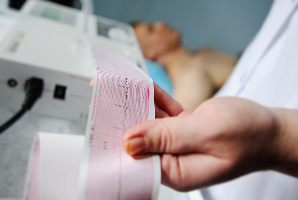
An ECG is painless and takes about ten minutes to perform. During the test, the nurse will attached electrodes (flat metal discs) to your arms, legs and chest. The electrodes are connected to a machine that records the electrical signals of each heartbeat. Every time your heart beats, it produces tiny electrical signals. An ECG machine records these signals onto paper, allowing your doctor or nurse to see how well your heart is functioning.
What does an ECG show?
The electrodes on the different parts of the body detect electrical impulses coming from different directions within the heart. There are normal patterns for each electrode. Various heart disorders produce abnormal patterns. The heart disorders that can be detected include:
Abnormal heart rhythms. If the heart rate is very fast, very slow, or irregular. There are various types of irregular heart rhythm with characteristic ECG patterns.
A heart attack (myocardial infarction), and if it was recent or some time ago. A heart attack causes damage to heart muscle, and heals with scar tissue. These can be detected by abnormal ECG patterns.
An enlarged heart. Basically, this causes bigger impulses than normal.